Difference between revisions of "LPD (lateralized periodic discharges)"
From EEGpedia
(Created page with "Synonyme: PLED (Periodic lateralized epileptiform discharges) *'''L'''ateralized: so unilateral *'''P'''eriodic: Relative constant interval between the discharges, which vari...") |
|||
| Line 3: | Line 3: | ||
*'''L'''ateralized: so unilateral | *'''L'''ateralized: so unilateral | ||
*'''P'''eriodic: Relative constant interval between the discharges, which varies between 0.5 and 3 seconds (most often around 1 second) | *'''P'''eriodic: Relative constant interval between the discharges, which varies between 0.5 and 3 seconds (most often around 1 second) | ||
| − | *'''D'''ischarge: Could be | + | *'''D'''ischarge: Could be [[Delta waves]] or sharp waves with a “epileptiform” morphology |
| + | |||
| + | |||
| + | * If a more ictal appearance it can be categorized as LPD+:<ref>ACNS STANDARDIZED ICU EEG NOMENCLATURE v. 2012</ref> | ||
| + | **'''+F''': superimposed fast activity. | ||
| + | **'''+R''': superimposed rhythmic or quasi-rhythmic activity. | ||
| + | **'''+FR''': superimposed fast activity and rhythmic or quasi-rhythmic activity. | ||
| Line 20: | Line 26: | ||
[[File:LPD%27s_left_parieto_occipital_male_78_yo_with_a_hemmorrhage_left_occipital_(average).png|border|1200px]] | [[File:LPD%27s_left_parieto_occipital_male_78_yo_with_a_hemmorrhage_left_occipital_(average).png|border|1200px]] | ||
---- | ---- | ||
| + | |||
| + | '''Notes''' | ||
| + | <references/> | ||
Revision as of 10:31, 19 May 2017
Synonyme: PLED (Periodic lateralized epileptiform discharges)
- Lateralized: so unilateral
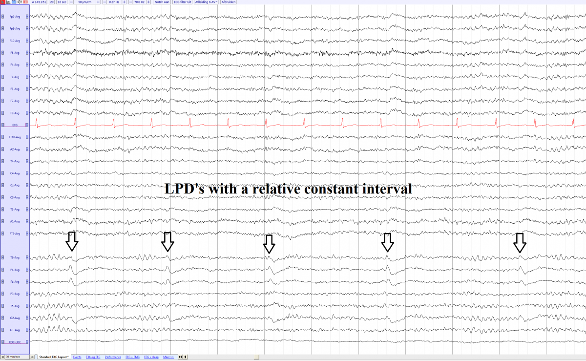
- Periodic: Relative constant interval between the discharges, which varies between 0.5 and 3 seconds (most often around 1 second)
- Discharge: Could be Delta waves or sharp waves with a “epileptiform” morphology
- If a more ictal appearance it can be categorized as LPD+:[1]
- +F: superimposed fast activity.
- +R: superimposed rhythmic or quasi-rhythmic activity.
- +FR: superimposed fast activity and rhythmic or quasi-rhythmic activity.
- LPD’s are seen in acute cerebral laesion
- Non specific:
- Acute cerebrovascular events
- Encephalitis
- Subdural hematoma
- Other structural lesions
- Non specific:
- Tend to disappear in weeks
- Higher risk of seizures in patients with LPD’s
LPD's left parieto occipital in a male of 78 years old with a hemmorrhage left occipital (average)

Notes
- Jump up ↑ ACNS STANDARDIZED ICU EEG NOMENCLATURE v. 2012